
Our Health Library information does not replace the advice of a doctor. Please be advised that this information is made available to assist our patients to learn more about their health. Our providers may not see and/or treat all topics found herein.
Kidney Stone Seen on Intravenous Pyelogram (IVP)
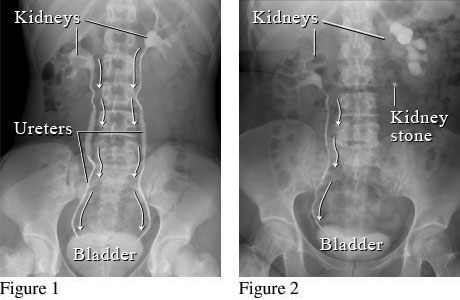
Courtesy of Intermountain Medical Imaging, Boise, Idaho.
These figures show an X-ray with contrast dye (intravenous pyelogram, or IVP) of the kidneys, ureters, and bladder. Figure 1 shows a normal flow from the kidneys, through the ureters, to the bladder (white arrows). Figure 2 shows a kidney stone blocking the normal flow of urine in the ureter on the right.
Current as of: July 26, 2023
Author: Healthwise Staff
Clinical Review Board
All Healthwise education is reviewed by a team that includes physicians, nurses, advanced practitioners, registered dieticians, and other healthcare professionals.


This information does not replace the advice of a doctor. Healthwise, Incorporated disclaims any warranty or liability for your use of this information. Your use of this information means that you agree to the Terms of Use and Privacy Policy. Learn how we develop our content.
To learn more about Healthwise, visit Healthwise.org.
© 1995-2024 Healthwise, Incorporated. Healthwise, Healthwise for every health decision, and the Healthwise logo are trademarks of Healthwise, Incorporated.